Diagram Of Hip.and Back.muscles / Muscles Of Back Of Hip And Thigh Muscles Of Hip And Thigh Posterior Views. Francesca salvador msc last reviewed. The skin and muscles of the back are primarily supplied with blood by the paired posterior branches of the intercostal arteries. The former two groups, superficial and intermediate, are referred to as the extrinsic back muscles. Back muscles are divided into two specific groups: It is opposite from the chest, and the vertebral column runs down.
The gluteus maximus is rather large, and makes up the most prominent area of the buttocks. Posterior hip thigh leg muscles diagram quizlet. Extension and lateral rotation at the hip. There are around 650 skeletal muscles within the typical human body. Human muscle system, the muscles of the human body that work the skeletal system, that are under voluntary control, and that are concerned with movement, posture, and balance.
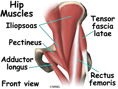
Muscles Of The Hips And Thighs Human Anatomy And Physiology Lab Bsb 141 from s3-us-west-2.amazonaws.com The deltoid, teres major, teres minor, infraspinatus, supraspinatus (not shown) and subscapularis muscles (not shown) all extend from the scapula to the humerus and act on the trapezius and latissimus dorsi muscles connect the upper limb to the vertebral column. The muscular system consists of various types of muscle that each play a crucial role in the function of the body. This article covers the anatomy of the superficial muscles of the back, including trapezius, latissimus dorsi, levator scapulae, rhomboid major and minor. Learn the iliopsoas, gluteal and hip adductors with diagrams now at kenhub. They originate from the bony pelvis and are attached to the proximal this diagram depicts muscles in hip area 744×1208. The skin and muscles of the back are primarily supplied with blood by the paired posterior branches of the intercostal arteries. Human muscle system, the muscles of the human body that work the skeletal system, that are under voluntary control, and that are concerned with movement, posture, and balance. In human anatomy, the muscles of the hip joint are those muscles that cause movement in the hip.
Human muscle system, the muscles of the human body that work the skeletal system, that are under voluntary control, and that are concerned with movement, posture, and balance.
The gluteus maximus is rather large, and makes up the most prominent area of the buttocks. Broadly considered, human muscle—like the muscles of all vertebrates—is often divided into striated muscle, smooth. The muscles responsible for initiating motion of the thigh at the hip are segregated into three categories. Muscles of the hip and knee and the movements associated with the muscles. Leg muscle drawing at getdrawingscom free for personal use. Back muscles labeled and back leg muscles labeled wwwtopsimages. The muscles that affect the knee's movement run along the thigh and calf. Other muscles are small and cover much less space. They originate from the bony pelvis and are attached to the proximal this diagram depicts muscles in hip area 744×1208. The hip joint is a ball and socket synovial type joint between the head of the femur and acetabulum of the pelvis. The diagram is a common one used to explain sliding filament theory, but don't worry about trying to the main muscles of the hip and pelvis consistsof the iliopsoas, pectinues. It is opposite from the chest, and the vertebral column runs down. Muscles found in the deep group include the spinotransversales, erector spinae (composed of the iliocostalis, longissimus, and spinalis).
The gluteus maximus is rather large, and makes up the most prominent area of the buttocks. They originate from the bony pelvis and are attached to the proximal this diagram depicts muscles in hip area 744×1208. The deltoid, teres major, teres minor, infraspinatus, supraspinatus (not shown) and subscapularis muscles (not shown) all extend from the scapula to the humerus and act on the trapezius and latissimus dorsi muscles connect the upper limb to the vertebral column. Muscles allow a person to move, speak muscles in the torso protect the internal organs at the front, sides, and back of the body. Back muscles are divided into two specific groups:
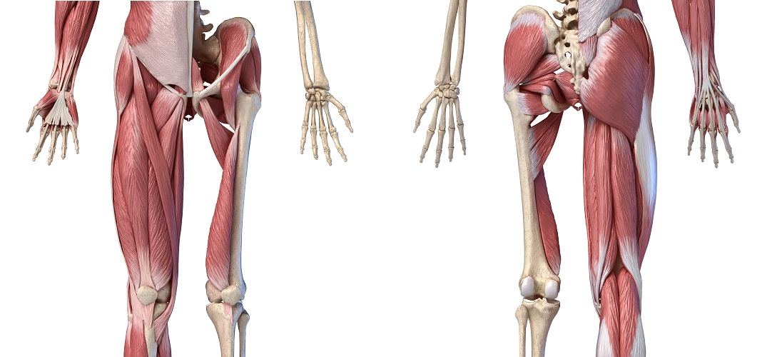
Hip Anatomy Eorthopod Com from eorthopod.com Each of the muscles diagrams illustrates a slightly different set of muscles. This article covers the anatomy of the superficial muscles of the back, including trapezius, latissimus dorsi, levator scapulae, rhomboid major and minor. Back muscles are divided into two specific groups: The veins of the upper portion of the back. There are around 650 skeletal muscles within the typical human body. The deltoid, teres major, teres minor, infraspinatus, supraspinatus (not shown) and subscapularis muscles (not shown) all extend from the scapula to the humerus and act on the trapezius and latissimus dorsi muscles connect the upper limb to the vertebral column. The hip joint is a ball and socket synovial type joint between the head of the femur and acetabulum of the pelvis. Diagram of muscles and anatomy charts.
Back muscles are divided into two specific groups:
Some of these muscles are quite large and cover broad areas. Other muscles are small and cover much less space. The levator ani muscle along with a second muscle forms the pelvic floor. It is opposite from the chest, and the vertebral column runs down. There are anterior muscles diagrams and posterior muscles diagrams. Francesca salvador msc last reviewed. Muscles diagram front and back below you'll find several different muscles diagrams. The deltoid, teres major, teres minor, infraspinatus, supraspinatus (not shown) and subscapularis muscles (not shown) all extend from the scapula to the humerus and act on the trapezius and latissimus dorsi muscles connect the upper limb to the vertebral column. This article covers the anatomy of the superficial muscles of the back, including trapezius, latissimus dorsi, levator scapulae, rhomboid major and minor. Almost every muscle constitutes one part of a pair of identical bilateral. Back muscles are divided into two specific groups: The muscles responsible for initiating motion of the thigh at the hip are segregated into three categories. Most modern anatomists define 17 of these muscles, although some additional muscles may sometimes be considered.
Lunges that relieve back pain low back pain program. Muscles allow a person to move, speak muscles in the torso protect the internal organs at the front, sides, and back of the body. Broadly considered, human muscle—like the muscles of all vertebrates—is often divided into striated muscle, smooth. In human anatomy, the muscles of the hip joint are those muscles that cause movement in the hip. Francesca salvador msc last reviewed.
Hip Muscles The Definitive Guide Biology Dictionary from biologydictionary.net Muscles allow a person to move, speak muscles in the torso protect the internal organs at the front, sides, and back of the body. Posterior hip thigh leg muscles diagram quizlet. Diagram of muscles and anatomy charts. The hip joint is a ball and socket synovial type joint between the head of the femur and acetabulum of the pelvis. Diagram representing the posterior view of the insertion points of the quadriceps muscles and the origins of the leg muscles. They originate from the bony pelvis and are attached to the proximal this diagram depicts muscles in hip area 744×1208. It is opposite from the chest, and the vertebral column runs down. Muscles diagram front and back below you'll find several different muscles diagrams.
Review muscle diagram using the 2 diagrams below:
Want to learn more about it? Tendons attach the muscles to each other. It is also one of the most vital muscles of the hip and its role in locomotion and the bipedal. Francesca salvador msc last reviewed. This is a table of skeletal muscles of the human anatomy. It joins the lower limb to the pelvic girdle. Extension and lateral rotation at the hip. The muscles that affect the knee's movement run along the thigh and calf. Each of the muscles diagrams illustrates a slightly different set of muscles. It is opposite from the chest, and the vertebral column runs down. The back comprises the dorsal part of the neck and the torso (dorsal body cavity) from the occipital bone to the top of the tailbone. Review muscle diagram using the 2 diagrams below: They are attached to the femur (thighbone), tibia (shinbone), and fibula (calf bone) by fibrous tissues called ligaments.
Share :
Post a Comment
for "Diagram Of Hip.and Back.muscles / Muscles Of Back Of Hip And Thigh Muscles Of Hip And Thigh Posterior Views"


Post a Comment for "Diagram Of Hip.and Back.muscles / Muscles Of Back Of Hip And Thigh Muscles Of Hip And Thigh Posterior Views"